Diagnostics post partum online course
Have you ever been in doubt when diagnosing an obstetric laceration?
What does a 2nd-degree perineal tear look like in real life? How about a 3rd- or 4th-degree tear? Correct perineal repair starts with the right diagnosis, so you need to know what you are looking for.
Get instant access to hours of free online teaching on diagnostics.
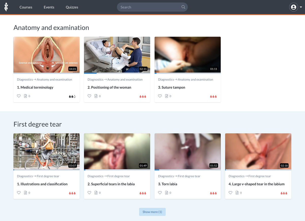
Perfect perineal repair? Start with the right diagnosis
Tearing is quite common in vaginal birth, and tears can occur anywhere in the vulva, the vagina, or the perineum. As clinicians, we aim to restore pelvic floor function in between and after childbirth.
In this course, you will learn how we use medical and technical terminology in connection with the anatomical structures to describe trauma after birth, and how symmetry and anatomical landmarks can assist your evaluation and diagnosis of the trauma. And why a rectal examination is an important part of the diagnostic process. The course includes:
- Animation videos and medical illustrations
- Training instructions with medical models
- Clinical videos and photos of patients
- Quiz with printable certificate available for Pro members – Activate your free account or subscribe to get full access.
See the full course overview here: https://my.gynzone.com/courses/64-diagnostics
All our clinical videos with patients are recorded with the written and oral consent of both patient and operator, in collaboration with the hospital.
Anatomy and examination

Get to know the superficial pelvic floor muscles and their functions, and get clinical tips on how to identify them in a laceration.
Positioning the woman and keeping her as comfortable and relaxed as possible is essential. If she wishes to nurse her newborn baby, she can be supported with a pillow under her elbow. It can be helpful to place the woman in the Trendelenburg position, in order to improve your access and visualization of the posterior wall of the vagina and the lower area of the perineum.
Many women prefer that information be provided with good eye contact, and possibly with a soft touch on the shoulder or a calming hand elsewhere on their body. Always remember that she may feel vulnerable and exposed in this situation.
Classification
Learn how to classify birth lacerations 1st to 4th degree, according to the RCOG guidelines
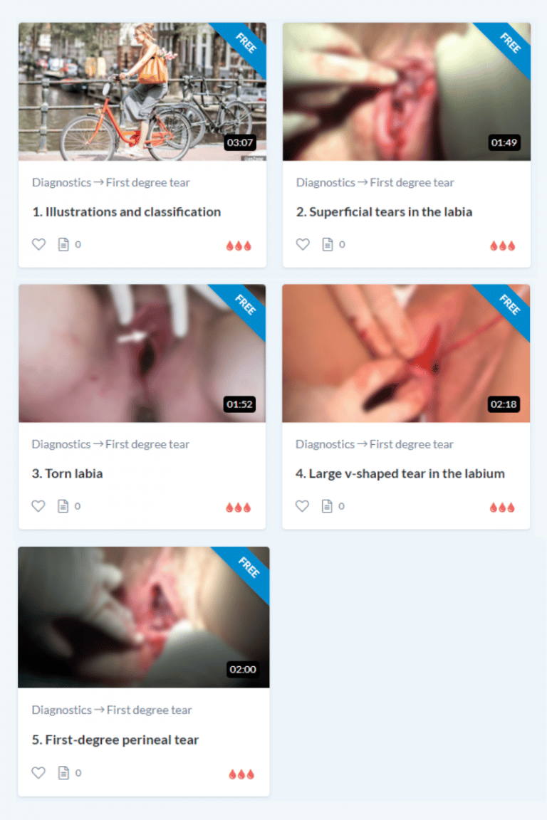
- First-degree tears, including labial lacerations
- Second-degree tears, involving the superficial perineal muscles
- Mediolateral episiotomy
- Grade 3 tears, involving the anal sphincters
- Grad 4 tears, involving the rectal mucosa
First-degree tears
First-degree tears are superficial tears in the perineal skin, the vaginal mucosa, around the hymen, and in the labia. The perineal muscles and fascia are not involved in first-degree tears. Superficial tears or skin abrasions or grazes can also occur on the inside of the labia. These tears may be sutured to minimize stinging pain during urination and to promote fast healing, or in order ensure hemostasis if the wound edges are bleeding.
Any bilateral internal tears on the labia should be sutured to prevent these tear edges from spontaneously healing across the vaginal opening or across the urethral opening. Some women report that if tears in the labia are not repaired, they have experienced pain and discomfort with intercourse, bicycling, or other direct pressure in the area later in life. Correct adaption of the tissue can also help to ensure a good cosmetic result so that the woman is satisfied with the appearance of the vulva and perineum following birth.
Second-degree perineal tears
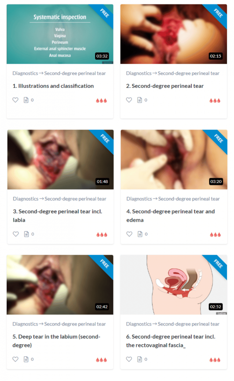
The rectovaginal fascia is a thin tissue structure lying between the vagina and the rectum. Its function is to hold the intestine in place.
Defects that involve the rectovaginal fascia can manifest as a rectocele later in life if they are left unsutured. The rectovaginal fascia can be recognized as a thin whitish structure. This video shows the rectovaginal fascia in a tear, where the vaginal mucosa is torn down to the fascia.
Episiotomy
The angle of an episiotomy is measured from the centerline, i.e. the line from the vagina down to the anus. An episiotomy at an angle of 0° is called a median episiotomy, or a midline episiotomy. An incision at an angle of 45 to 60 degrees starting from the introitus is called a mediolateral episiotomy.
The recommended angle is 60 degrees so that the scissors are angled out towards the woman’s thigh and away from the anal sphincter muscles.
An episiotomy at an angle of 60 degrees starting from 1-2 cm over the introitus is called a lateral episiotomy.
Midwives and doctors in some countries prefer to make the incision on the woman’s right side.
In other countries, an incision towards the woman’s left side is recommended. The same anatomical structures are involved in each case.
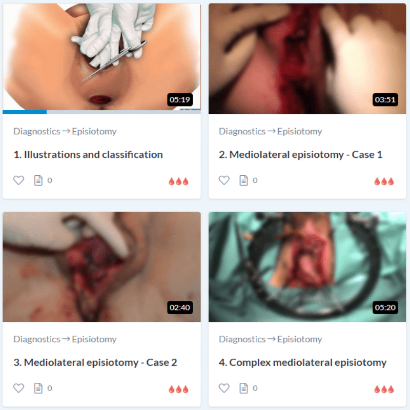
1. Perineal skin
2. Vaginal mucosa
3. Bulbocavernosus muscle
4. Poss. Transverse perineal muscle
An episiotomy can involve everything from a superficial incision in the skin to a very extensive cut in the woman’s perineum.
The prerequisite for a correct diagnosis is that you perform a systematic inspection of the vulva, vagina, perineum, the external anal sphincter muscle, and the anal mucosa. Rectal examination is always necessary in order to determine whether the external anal sphincter muscle or the anal mucosa are involved in the tear beyond the episiotomy.
Third-degree perineal tears
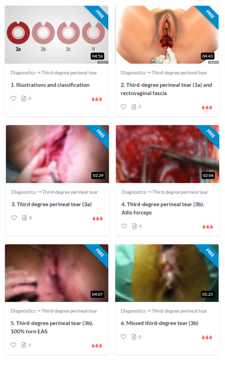
In the professional literature and guidelines, tears of this type are described as OASI. This is an abbreviation for Obstetric Anal Sphincter Injury.
The external anal sphincter muscle is abbreviated EAS.
The internal anal sphincter muscle is abbreviated IAS.
Tears are defined as degree 3a, 3b or 3c depending on how much of the musculature is involved.
Fourth-degree perineal tears
The WHO ICD diagnosis codes are used to document diagnosis in medical records for international comparisons.
ICD11 codes
Codes DO070.0 to DO070.9 are generally used for perineal lacerations during delivery.
Code DO70.0 is used for first-degree tears. More precise codes have also been developed to indicate the location of the tear.
DO70.0A is a first-degree tear in the vulva. The vulva is the area surrounding the woman’s entire genital opening, including the area around the clitoris, labia, introitus, hymen and perineum.
DO70.0B is a first-degree tear in the vagina. The vagina is defined as the area extending from the hymen in towards the cervix.
DO70.0C is a first-degree tear in the perineal skin. This skin covers the area from the introitus down to the anus.
DO70.0D is a first-degree tear of the labia. The labia surround the vaginal opening.
DO70.1 is used for second-degree tears, involving the bulbocavernosus muscle and/or the transverse perineal muscle.
DO70.1B is used if there is also a tear in the rectovaginal fascia or septum rectovaginalis.
DO70.2” is used for third-degree tears.
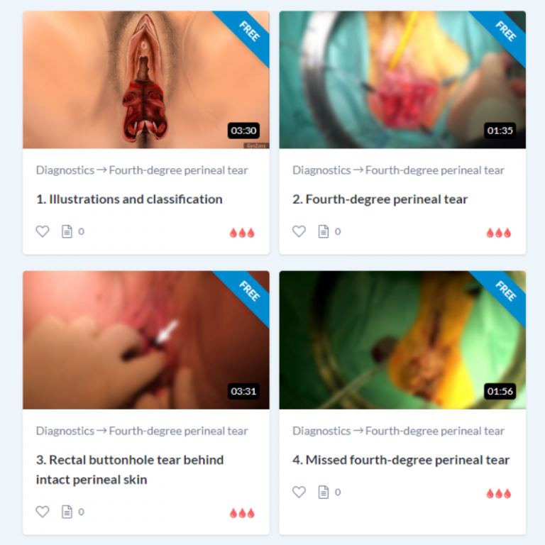
DO70.3 is used for fourth-degree tears.
DO70.4 is used if an isolated tear has formed between the rectum and vagina, but does not involve the anal sphincter complex. This is also known as a buttonhole defect and can occur even behind intact perineal skin, as seen in this video.
That’s what users are saying
Authors of the course

Sara Kindberg
Midwife, PhD, Founder of GynZone Clinical Perineal Care Specialist.
Sara wrote her PhD Thesis on suturing of second-degree perineal trauma in 2008.

Karl Møller Bek
Urogynecologist, PhD, Founder of GynZone Scandinavian specialist in the reconstruction of anal sphincter injuries Karl wrote his PhD Thesis on the diagnosis and treatment of anal sphincter trauma in 1993.

Peggy Seehafer
Midwife, Anthropologist Peggy has 30 years of clinical experience, and is editor of the German Journal of Midwifery.

Marianne Glavind-Kristensen
Urogynecologist, PhD Senior Consultant and Head of Pelvic Floor Unit at Aarhus University Hospital Marianne wrote her PhD Thesis in 2001.


