
RESOURCES
Perineal tear classification: 1st to 4th degree
How does perineal tear classification work, from first to fourth degree?
Perineal tear classification grades a birth injury by the deepest structure it reaches, from the perineal skin down to the anorectal mucosa. Most units follow the RCOG system, which runs from first to fourth degree and splits third-degree tears into 3a, 3b, and 3c. The degree of tear you diagnose determines who repairs it, where the repair takes place and how the woman is followed up. An accurate diagnosis depends on a systematic examination, including a rectal examination, that identifies the deepest structure involved.
This guide covers the landmarks, the rectal examination, and how to separate a second-degree tear from a third-degree tear.
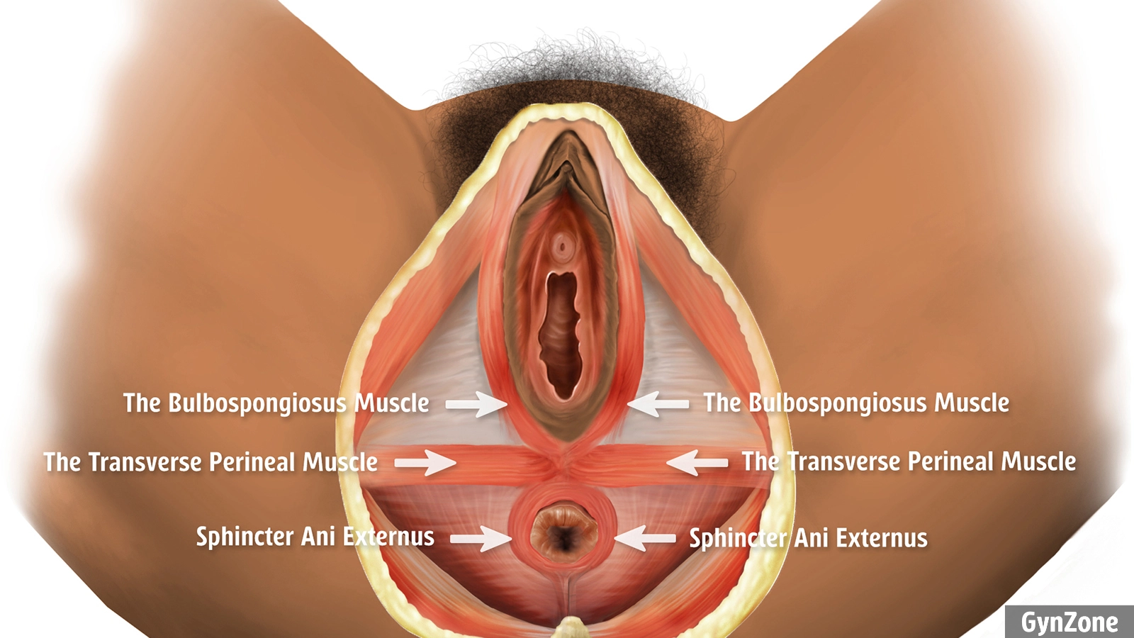
The superficial perineal muscles and the external anal sphincter, which sits just beneath the perineal skin. Illustration from the GynZone diagnostics course.
The RCOG classification of perineal tears at a glance
RCOG Green-top Guideline No. 29 uses the classification described by Sultan. Each degree is defined by the anatomy involved. The length of the tear doesn’t count. ICD-11 codes all four degrees under JB09, and lists types 3a, 3b, and 3c as index terms under JB09.2, so write the subtype in your notes as well as the code.
| Degree | Structures involved (RCOG) | ICD-11 | ICD-10 |
| First degree | Perineal skin and/or vaginal mucosa | JB09.0 | O70.0 |
| Second degree | Perineal muscles, anal sphincter intact | JB09.1 | O70.1 |
| Third degree, 3a | Less than 50% of external anal sphincter (EAS) thickness torn | JB09.2 | O70.2 |
| Third degree, 3b | More than 50% of EAS thickness torn | JB09.2 | O70.2 |
| Third degree, 3c | Both EAS and internal anal sphincter (IAS) torn | JB09.2 | O70.2 |
| Fourth degree | EAS, IAS, and anorectal mucosa torn | JB09.3 | O70.3 |
| Rectal buttonhole tear | Anorectal mucosa torn, anal sphincter complex intact | Not a fourth-degree tear, document it separately | Follow your national coding |
Third- and fourth-degree tears together are obstetric anal sphincter injuries (OASI). In the UK, RCOG reports an incidence of 6.1% in first births and 1.7% in women who have given birth before.
Anatomical landmarks to find on examination
Symmetry and a few fixed landmarks give you an overview before you count layers.
- The hymenal remnant. Violet and often swollen after birth, about 1 cm inside the introitus. It marks the border between the introitus and the vagina.
- The apex of the vaginal tear. Always locate it. Your repair starts above it.
- The bulbospongiosus muscle. It surrounds the vaginal opening. Torn ends retract up along the labia majora.
- The transverse perineal muscle. It runs across the perineum. Torn ends retract out to the sides.
- The external anal sphincter (EAS). Striated, dark red (often compared to raw beef), roughly 1 cm thick, extending about 3 cm up along the anal canal, just beneath the perineal skin surrounding the anus.
- The internal anal sphincter (IAS). Smooth muscle, whitish (often compared with chicken meat), a few millimetres thick, lying inside the EAS.
- The rectovaginal fascia. A thin whitish layer of strong connective tissue between the vagina and the rectum.
First degree means skin, hymen or mucosa only – no muscle fibres
Superficial lacerations of the vulva that may involve the labia, vaginal mucosa, hymen, and perineal skin, but no muscle or fascia. The reasons to suture a first degree tear are to restore the anatomy and give the woman a good functional and cosmetic result. If a vessel is bleeding, it may need ligating. Always suture bilateral labial tears: otherwise the wound edges can heal together across the vaginal opening or over the urethra.
Second degree involves the perineal muscles
One or both superficial perineal muscles are torn, and the anal sphincter is intact. The tear often opens in a heart shape as the muscle ends of the bulbospongiosus retract upwards, towards the labia majora. A tear in the perimysium, around the external anal sphincter muscle, is also classified as a second-degree tear. An episiotomy is usually equivalent to a second-degree perineal tear. Watch for lateral tears, tears behind intact perineal skin, and rectovaginal fascia defects, which can lead to a rectocele if left unrepaired. Our tips for assessing a second-degree tear go further.
Third degree reaches the anal sphincter complex (OASI)
A third-degree tear involves the EAS and possibly also the IAS. The subtype (3a-c) depends on how much of the EAS thickness is torn, and whether the IAS is involved.
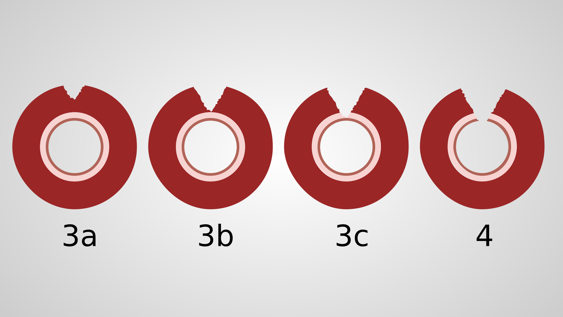
Cross-section of the anal sphincter complex. The wedge shows how deep each grade of OASI reaches.
Fourth degree reaches the anorectal mucosa (OASI)
Both sphincters and the anorectal mucosa are torn. With your finger in the anus, you see your glove where the tissue between the vagina and rectum should be.
Learn from real-world clinical cases through detailed videos.
The rectal examination comes with every classification
RCOG states that every woman with a vaginal birth is at risk of OASI, and that sphincter and anorectal mucosal injury cannot be excluded without a rectal examination. Examine systematically before you pick up a needle. One observational study cited by RCOG found that increased vigilance can double the detection rate.
- Explain what you’ll do, get her consent, and check that her analgesia works.
- Insert your index finger into the anus and place your thumb on the perineum.
- Palpate the EAS from 9 o’clock to 3 o’clock, so a defect slightly off the midline isn’t missed.
- Ask her to contract around your finger, and lift the rectal mucosa to inspect the posterior vaginal wall.
Second or third degree? How to tell them apart
Take your time with this call. In our course material, a woman with a missed 3b tear came back three years after birth with flatus and faecal incontinence.
- Intact skin over the anus doesn’t rule out OASI. A 3a lesion can sit in the middle of the EAS and only show up on palpation.
- A torn perimysium is not a third-degree tear. When the whitish capsule or sheath is torn over an intact EAS, the tear stays second degree.
- Feel for lost substance. A hollow on one side, or thin tissue between your finger and thumb, suggests the sphincter ends have retracted.
- If in doubt, grade upwards. RCOG advises classifying an uncertain 3a/3b tear as 3b, and calling a more experienced colleague when the extent is unclear.
After the repair, examine again
After the repair, do a rectal examination, palpate per rectum and feel for stitches. RCOG recommends removing any suture you find through the anorectal mucosa, unless it is part of a fourth-degree repair, because a misplaced suture can lead to a rectovaginal fistula.
See every degree on real patient cases
Definitions only take you so far. Intact perineal skin can hide a second-degree tear or a buttonhole defect, and some lesions only show up under your fingers. In the GynZone diagnostics course, we take you through clinical videos and photos of first- to fourth-degree tears, including missed tears and buttonhole defects, with consent from every woman filmed. When you diagnose an OASI, the OASI repair course picks up where diagnosis ends.
Frequently asked questions
What are the different degrees of tearing during childbirth?
There are four. A first-degree tear involves only the perineal skin, hymen or vaginal mucosa. A second-degree tear involves the perineal muscles, with the anal sphincter intact. A third-degree tear reaches the anal sphincter complex and is split into 3a, 3b, and 3c by how much of the sphincter is torn. A fourth-degree tear also goes through the anorectal mucosa.
What is a 4th degree perineal laceration?
A fourth-degree tear involves the external anal sphincter, the internal anal sphincter, and the anorectal mucosa. It’s coded JB09.3 in ICD-11 and O70.3 in ICD-10. If the anorectal mucosa is torn but the sphincter complex is intact, it’s a rectal buttonhole tear, not a fourth-degree tear, and you document it separately.
How common are third- and fourth-degree tears?
RCOG reports an overall incidence of 2.9% in the UK, with a reported range of 0 to 8%. The rate is 6.1% in first births and 1.7% in women who have given birth before. Detection depends on the examination, so a low rate in your unit isn’t necessarily reassuring.
What is the difference between a perineal tear and an episiotomy?
A perineal tear happens spontaneously during birth. An episiotomy is a cut you make to enlarge the vaginal opening. A mediolateral episiotomy usually involves the same muscles as a second-degree tear, but it can extend into the anal sphincter, so you classify it with the same rectal examination. Our course in episiotomy repair shows how to repair one.
Do you need to suture a first-degree tear?
Not always. NICE advises suturing first-degree trauma to improve healing, unless the skin edges are already well approximated. Even when the edges are well approximated, two situations still need treatment. Ligate any vessel that is still bleeding. And always suture bilateral labial tears, because the edges can heal together across the urethral or vaginal opening.
References
- Royal College of Obstetricians and Gynaecologists. The management of third- and fourth-degree perineal tears. Green-top Guideline No. 29. 2015. https://www.rcog.org.uk/media/5jeb5hzu/gtg-29.pdf
- Sultan AH. Obstetric perineal injury and anal incontinence. Clinical Risk. 1999;5:193–6.
- National Institute for Health and Care Excellence. Intrapartum care (NG235). 2023. https://www.nice.org.uk/guidance/ng235/chapter/Recommendations
- World Health Organization. ICD-11 for Mortality and Morbidity Statistics, JB09 Perineal laceration during delivery (JB09.0 to JB09.3).